This brain mask atlas was created from the thresholding of the MNI 152 brain mask. The cerebrospinal fluid (CSF) spaces innaccuracies were corrected by an expert to properly represent the brain anatomy (e.g. the sepation between the frontal and temporal operculum). More precisely, this brain mask was used to enhance the accuracy of virtual glioma growth patterns.
Notice
If you use this atlas, please reference the corresponding article:Expert-validated CSF segmentation of MNI atlas enhances accuracy of virtual glioma growth patterns. A Amelot, E Stretton, H Delingette, N Ayache, S Froelich, and E Mandonnet. Journal of Neuro-Oncology, 2014. PDF.
Available Data for this Atlas:
- Brain Mask (147 Ko). This is a nifty image of the expert revised white and grey matter.
- White Matter Mask (122 Ko). This is a nifty image of the expert revised white mask tensorial image.


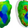
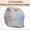 Non invasive cardiac personalisation
 Simulation of ventricular tachycardia re-entry circuit
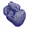 heartMeshFine
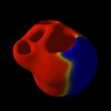 Electrophysiology
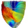 Cardiac Fibres from in vivo Diffusion Tensor Imaging
