


What are the main mechanisms involved in an asthma crisis? How do curative aerosols or the dust pollution deposit in the airways? How do emphysema or fibrosis affect the supply of air?
These are some of the questions we would like to contribute to answer. Our purpose is then to simulate the respiratory system from the ventilation process, and the mechanical behavior of the lung to the aerosol deposition.
- Ventilation air air flow modelling
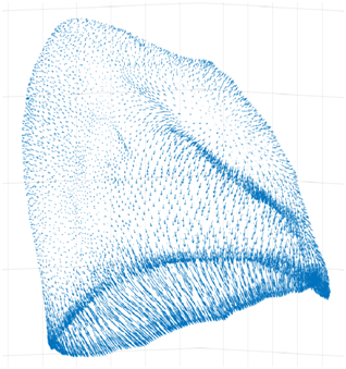
We simulate the air flow in the proximal part (till the 4th—6th generation) of a human bronchial tree on a real geometry coming from CT Scan.
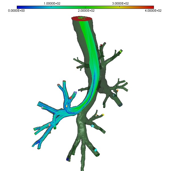
Streamlines at expiratory peak
The downstream part is taken into account by reduced lumped models (linear or nonlinear) describing the resistives small airways and the elastic recoil of the lung tissue, the whole system being driven by the external pleural pressure.
From these simulations we can obtain spirometric curves and try to reproduce pathological states.
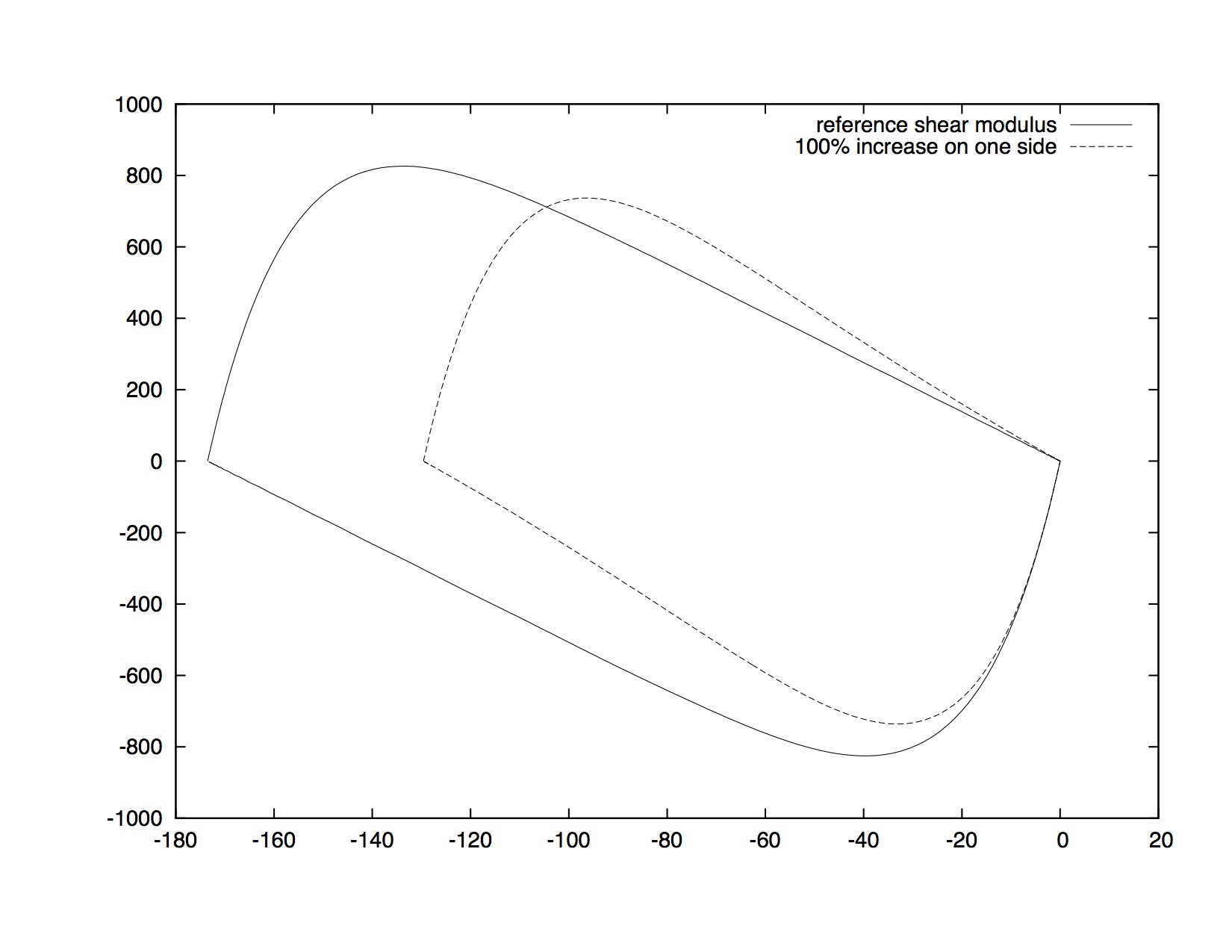
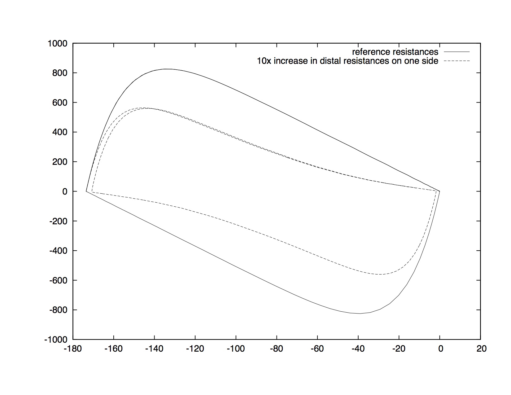
Spirometric Volume-Flow rate curves for different values of the lung elasticity
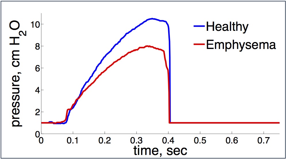
Measured pressure at the trachea for healthy or emphyema rats
Air velocity field in rat airways
The same kind of model is applied to the mechanical ventilation of healthy tats and rats with emphysema, where the parameters of the model are fitted with experimental mesurements. The aim is to develop an in-silico framework to better understand how aerodynamics changes in emphysema.
Collaboration with University California in San Diego (UCSD), USA
J. Oakes, A. Marsden and C. Darquenne
- Lung tissue modelling
We model the mechanical behavior of the lung parenchyma and derive several viscoelastic models.
We obtain a model for the lung tissue irrigated and supplied with air by a resistive bronchial tree. It is a viscoleastic non local (both in time and space) model. We recover some normal and pathological spirometric curves by choosing heterogenous elastic and resistive parameters.
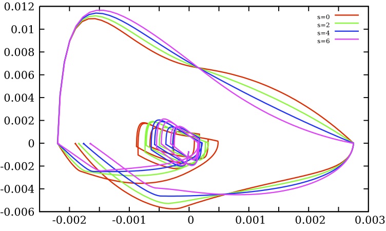
We also derive a viscoelastic model that reproduces the sound wave absorption for a certain range of frequencies of the lung parenchyma.
- Transport of aerosol
Our simulations are based on a fluid/kinetic coupling. Among other physical phenomena, in a joint work with experimentalists from INSERM Tours, we studied the transport of aerosol in an endotracheal tube (ETT) and recovered part of the deposition topography.
Simulated aerosol deposition
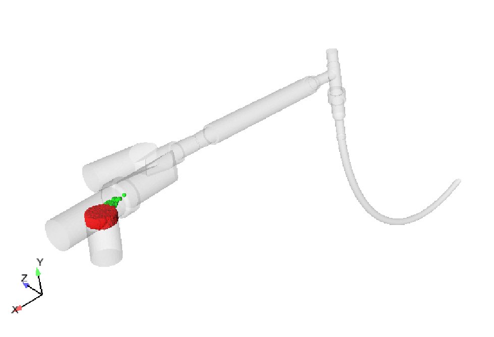
Launch of particles in an ETT device.
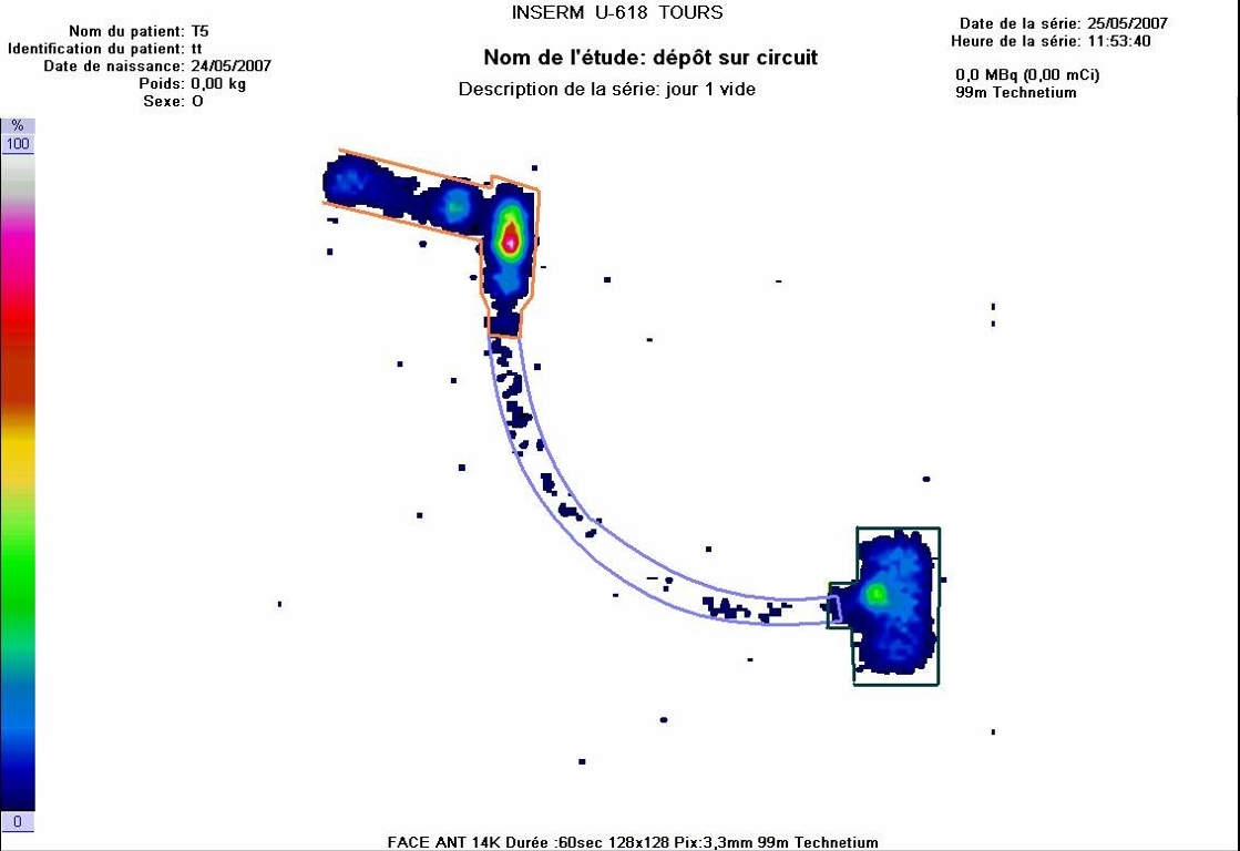
Measurements
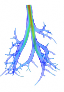
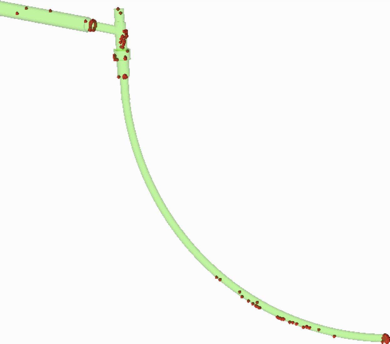
In collaboration with UCSD we also simulate the particule deposition in a rat bronchial tree at inspiration. The goal is to estimate the repartition of aerosol in each lobe, and how a disease such as emphysema affects this deposition.
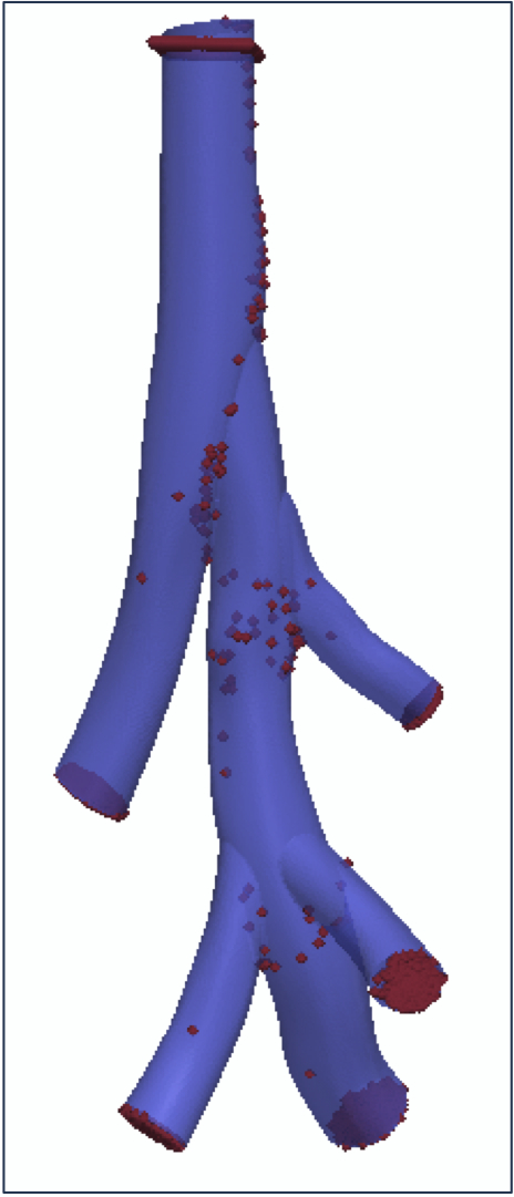
Particle deposition in a rat bronchial tree
- Tree-parenchyma coupled model for lung ventilation
We propose a lung ventilation model in which the parenchyma is described as a 3D homogenized elastic medium. It is strongly coupled to a 0D tree model. The tree induces an extra viscous term in the system constitutive relation. Geometries are exracted from computed tomography images. Surface displacement boundary conditions registered from images can be applied as boundary conditions The model is used to simulate healthy cases and pathological configurations where the parenchyma mechanical properties are altered or airways are constricted. Physiologically relevant behaviors are recovered.
Based on this model, the efficiency of treatments such as low density helium-oxygen mixtures can be investigated. This model is also used along with machine learning techniques to diagnose bronchoconstrictions from the analysis of lung ventilation dynamic images.

Cut of a simulation mesh (each terminal tree airway irrigates a given parenchyma region)
Left lung surface displacement, extracted from medical images
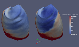
Simulated lung ventilation in a healthy case and when an airway is constricted