


Heart deseases are one of the main death causes and the study of cardiac electrophysiology is of current interest from medical and mathematical point of view. Our purpose is to simulate the electrical activity of the heart. In particular, we are interested in providing realistic healthy simulations and Electrocardiograms and in reproducing some common pathologies such as infarctions, bundle branch blocks and atrioventricular blocks.
- Simulations of the propagation of the action potential in the heart
Our simulations are performed with a mathematical model based on partial differential equations, our reference model is the bidomain model. The geometry we consider is a realistic heart mesh in which the atria surface is included. Important cardiac features are modeled: fibers anisotropy, cell heterogeneity (different action potential durations in the left ventricle) and fast-conducting networks.
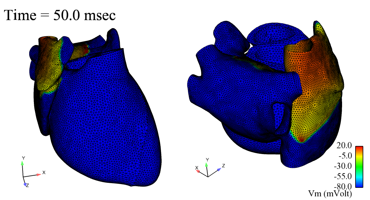
Action potentials during atrial depolarization
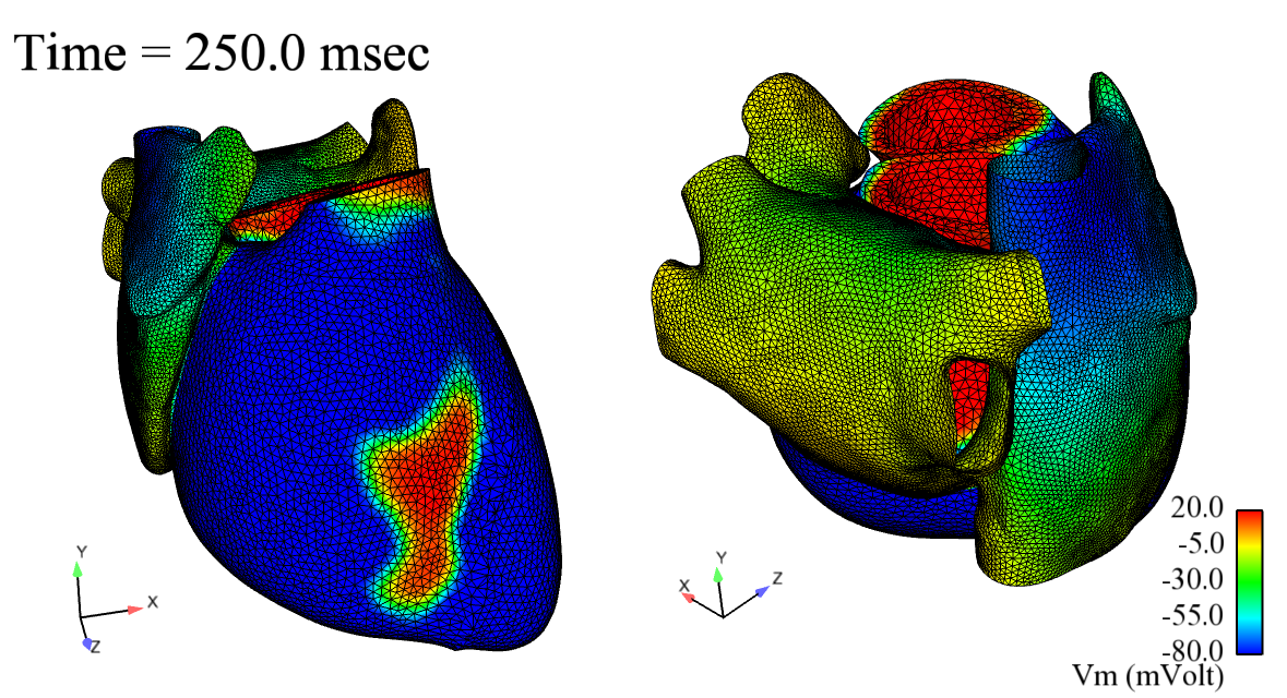
Action potentials during ventricular depolarization
- Simulation of electrocardiograms

Body surface during ventricular depolarization
Electrocardiogram is the most used exam providing an interpretation of the heart’s electrical activity. It records by skin electrodes the voltage in different body’s points, these measures (or leads) are then represented as twelve graphs of the recorded voltage vs time.
From a numerical point of view, to get electrocardiograms, the electrical activity of the heart is coupled with the electrical activity of the exterior medium, the human body. As the atrial surface is included in our model we obtain realistic P-wave complete electrocardiograms.
Electrocardiograms can detect the most of the known heart pathologies. Our purpose is to reproduce the most common ones and to obtain the associated Electrocardiograms. Good results have been obtained concerning for instance infarctions, Bundle Brunch Blocks and atrioventricular blocks.

Simulated 12-leads Electrocardiograms: healthy case (black) and a Left Bundle Brunch Block (blue)
- Reduced-order modeling
In order to decrease the computational cost of the simulations we implemented some reduced-order models.
For instance, a classical method that we use is the Proper Orthogonal Decomposition (POD). The accuracy of the reduced-order model has been assessed through electrocardiograms and it has been adapted in the case of some pathologies such as infarctions. In fact, a standard POD basis is not accurate enough to simulate this common cardiac desease so that the implementation of an “enlarged” POD basis has been necessary.

A basis from “modified” Schrodinger operator
We are investigating as well a new approach wich implies the evolution in time of basis. This new technique has been called ALP (from Approximated Lax Pairs) and first applied to cardiovascular system. On the key point of the method is the choice of the initial basis, we show here for instance a basis obtained from a “modified” Schrodinger operator, including the heart fibers orientation.
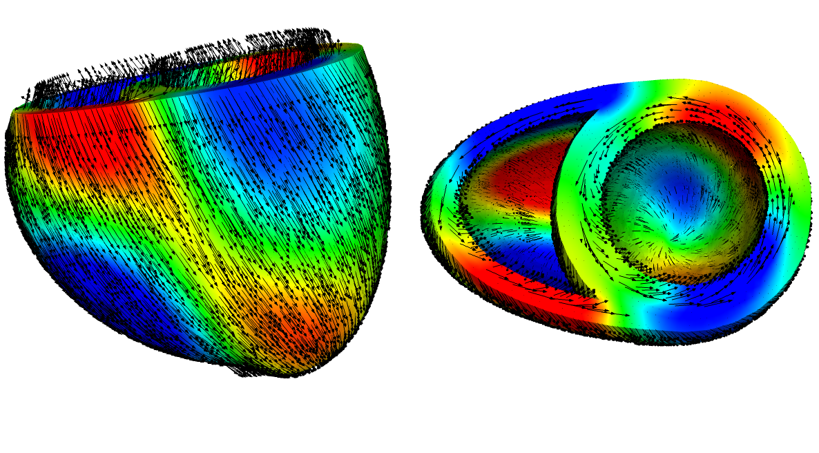
A basis from “modified” Schrodinger operator
- Inverse problem in electrocardiology

Snapshots of reference solution (left) simulated with a complete model and of inverse problem solution (right) simulated with POD method.
We are interested as well in inverse problems. We concentrate in particular on parameters identification, i.e. given a reference Electrocardiogram we want to deduce the model parameters useful to reproduce it. In order to solve this minimization problem we use an evolutionary algorithm and we apply a reduced-order model.
An exemple of application consist of identify infarction locations from synthetic electrocardiograms. In order to solve this problem we apply a Modified Genetic Algorithm and we use a “enlarged” POD basis.