


Website : http://nurea-soft.com
Nurea designs softwares simplifying surgeon decision process and patient follow-up. By automatizing medical images treatment and using powerfull algorithms developed during more than 10 years of research, Nurea is able to provide new relevant information to the surgeons. The software is completely integrated in the surgeon workflow making its use simple and intuitive with no effort. Thanks to our software, the surgeon saves time and the patient follow-up is enhanced. We first focus on aortic aneurism. The software allows the surgeon to easily visualize the evolution of the aneurysm through geometrical data but also pressures and stress due to the blood flow. We also integrate in our software, a prediction tool giving hints on how the aneurysm will evolve based on data classification of geometry and blood flow characteristics. The software is simple to use, intuitive and does not require any particular expertise in image processing, mathematics or modelisation such as other softwares on the market. The only task performed by the user is the loading of the images. All the information are then delivered automatically.
This start up is lead by Florian Bernard, who has an engineering degree from Enseirb-Matmeca. He gets a PhD in fluid dynamics from the mechanical and aerospace engineering department of Politecnico di Torino and a PhD in applied mathematics and scientific computing from University of Bordeaux.
Automatic Segmentation
In February 2019 Memphis gets funding from AMIES (Agence pour les mathématiques en intéraction avec l’entreprise et la société) for a one-year contract engineer in close collaboration with Nurea. His work consists in developing automatic segmentation of aortic aneurism.
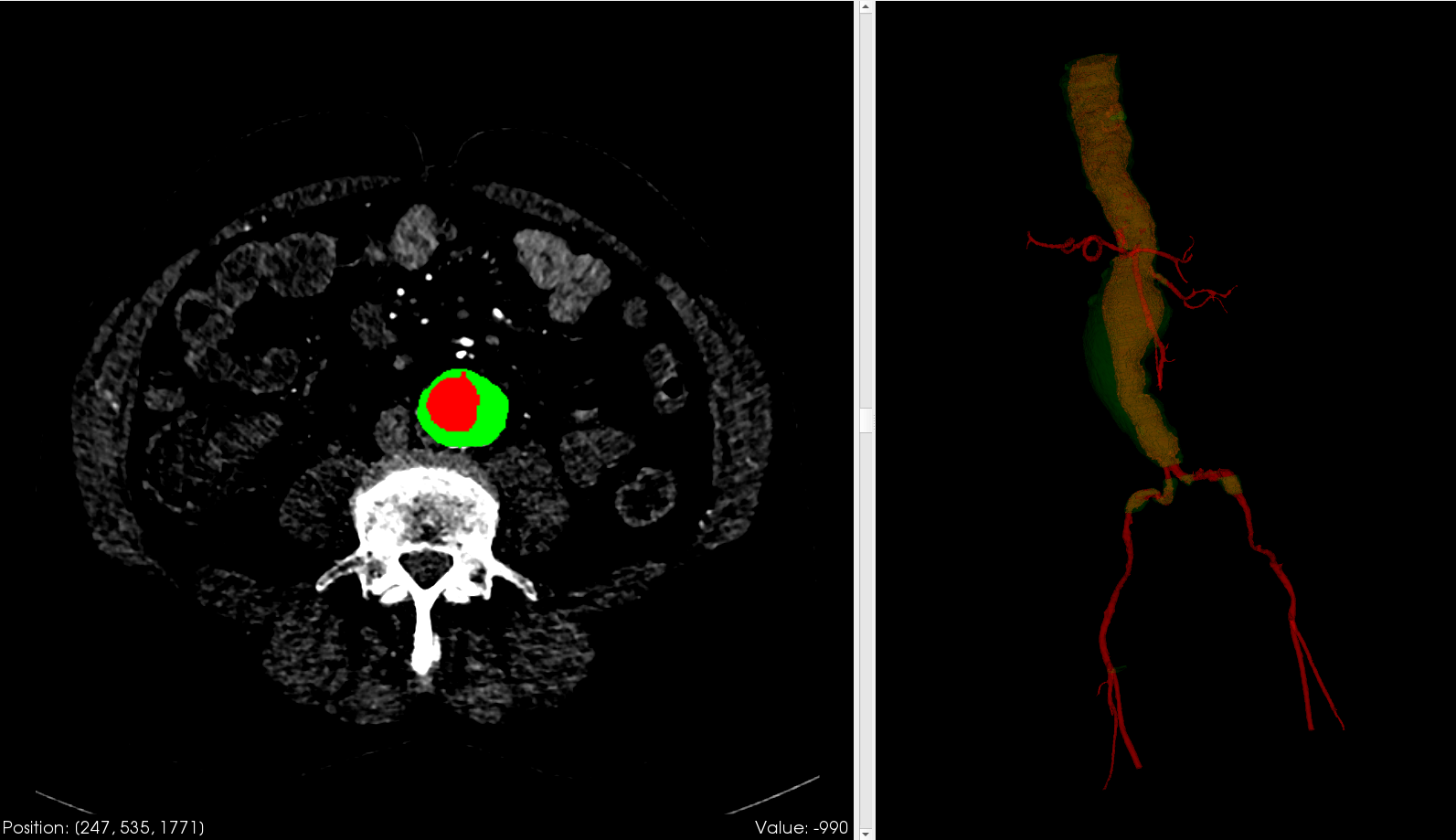
Example of aortic aneurism segmentation. Left: CT scan image. Right: 3D visualization. Red: blood; green: aortic wall
Registration
A method was developed to perform the registration of 2 segmentations from the same patient, obtained at different times. The objective was to compare the 2 segmentations and see how and where the aneurism has evolved. The method relies on the Iterative Closest Point algorithm and consider the outer surface of the aneurism. The registration is performed on the portion of the aorta starting right below the aortic bifurcation and ending right above the renal arteries.
In the following example, we register 3 segmentations on the one obtained from the post-operative control scan. We can see that the aneurism had grown uniformly between 2019 and 2020.

White: first post-operative segmentation. Red: registered segmentation
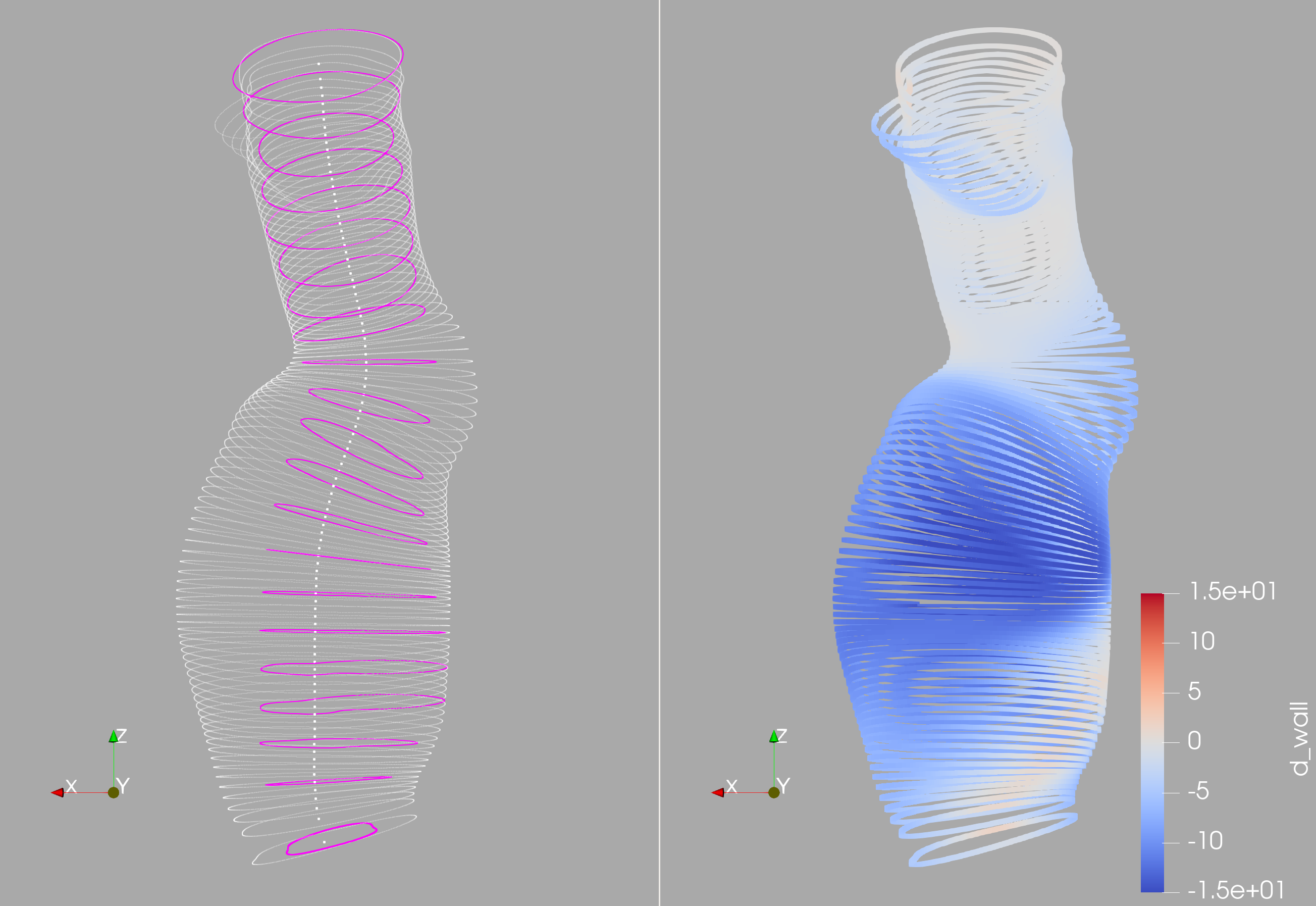
An other way to see the evolution is to look at the displacement field:
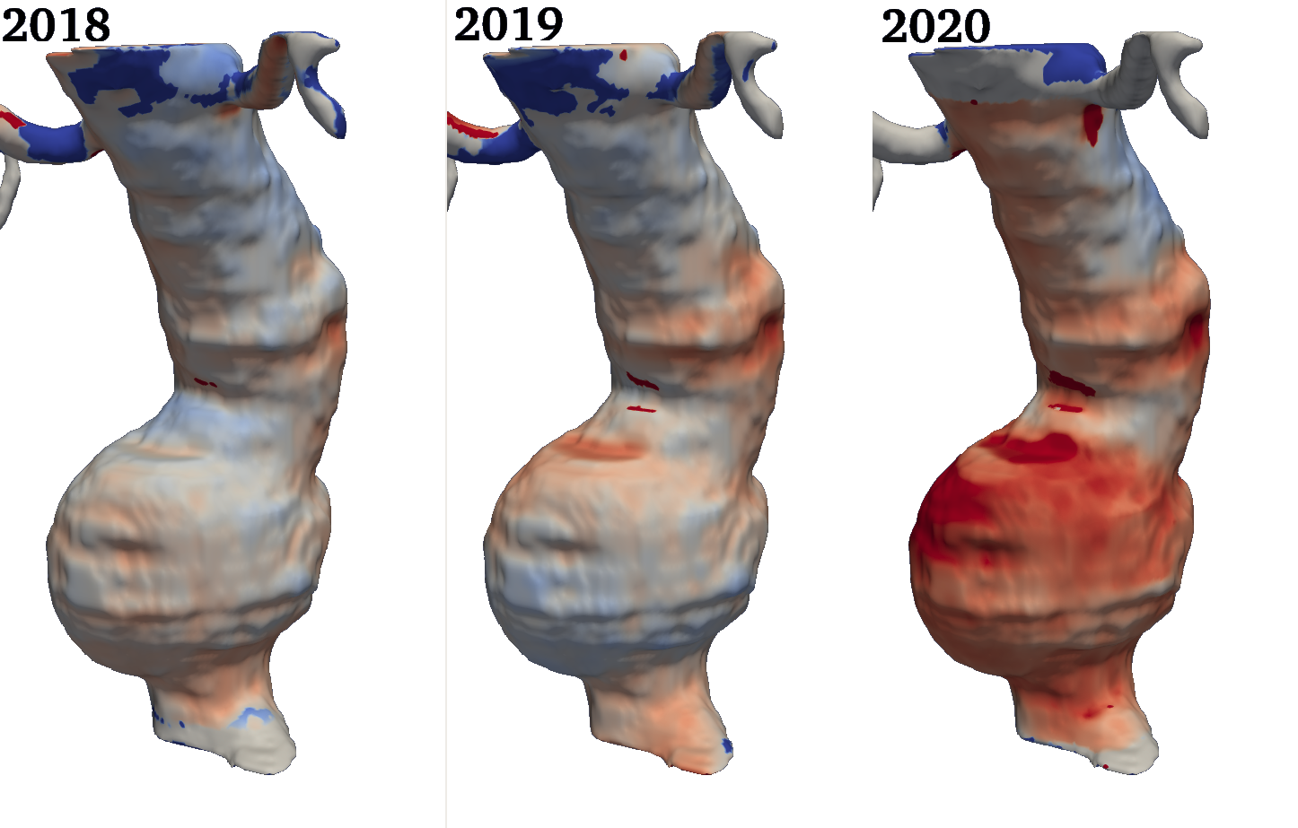
White: the displacement is null. Blue: the displacement is negative (the aneurism has shrank). Red: the displacement is positive (the aneurism has grown)
Here is an example of an aneurism that has shrunk after 2 years:

White: first post-operative segmentation. Red: registered segmentation
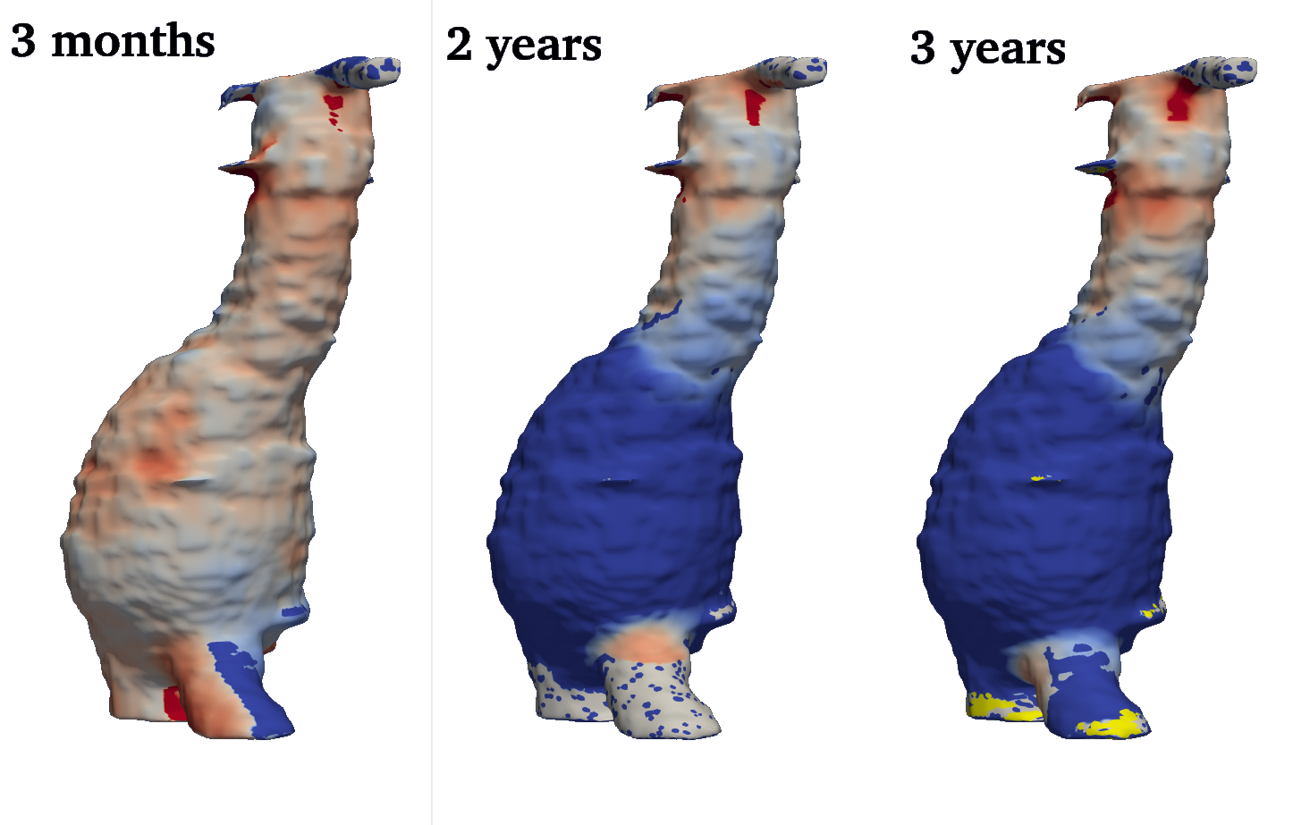
White: the displacement is null. Blue: the displacement is negative (the aneurism has shrank). Red: the displacement is positive (the aneurism has grown)
For more information on this start up do not hesitate to send an email at : contact@nurea-soft.com
Lauréat Concours d’innovation Ilab 2019
Parametrisation
Abdominal aortic aneurysm is a complex geometry: when performing analysis and observation we are faced with the peculiarities of the shape of the aneurysm sac, its deformations in proximity of the iliac bifurcation, and eventual bumps and irregularities given either by the recirculation processes that take place inside the sac, or by some imperfections in the acquisition of the CT scans or in the reconstruction process.
Therefore there is the need of reading the initial shape in a simple way, introducing some simplifications that allow the analysis of different aneurysms, presenting them in a more synthetic way. Moreover comparison between different patients must rely on some shared parameters over two or more geometries.
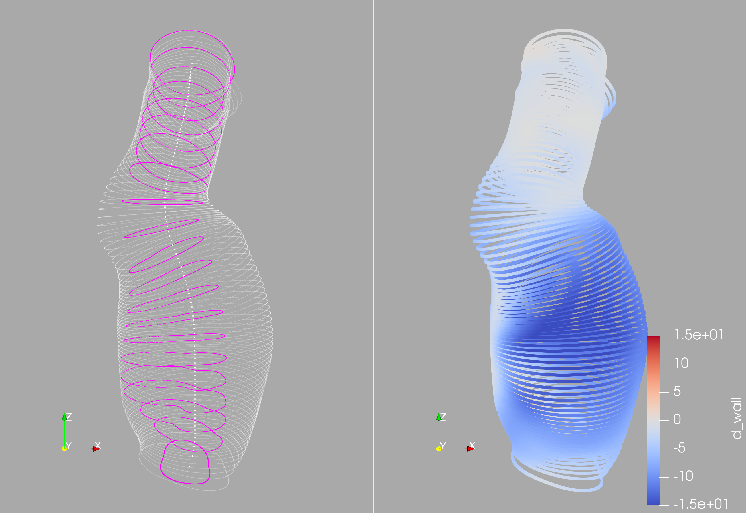
In this context we have elaborated a parametrisation procedure that starting from the 3D reconstruction of the aorta, isolates the trait of the aorta between the renal arteries and the iliac bifurcation, getting rid of the spurious blood vessels that might appear in the reconstruction and therefore leaving only the aneurysm sac.
Starting from the automatic segmentation, the parametrisation follows a pipeline based on the individuation of the centreline of the main vessel first, and then on the reconstruction of the aortic wall thought the selection of some section, individuated on the planes perpendicular to the centreline. Each section profile is parametrised via Fourier series, allowing its section to be defined solely by Fourier coefficients.
Below an example of some parametrised aneurysm and a few details about the parametrisation procedure here Parametrisation details.

Initial aneurysms shapes with the relative parametrisation. In red the centreline, in black the profile of the sections used to parametrise the aortic wall.
Parametrisation allow the quick confrontation and handle of the aneurysm shape. In particular we can now represent the aneurysm only by it centreline and the Fourier coefficients related to all its sections.
Registration.
As first ground to assess the validity and convenience of the parametrisaion, we briefly present a preliminary result for registration.
As the parametrisation allows us to define a common geometrical structure for all the aneurysms, it’s now easily compare them. Before presenting the result we list the hypotheses of wrk we have been following: when comparing two aneurysms, we consider that no torsion has occurred in the aorta, and that the extension, if present, is negligible, and finally we take the renal arteries as fixed point. These hypotheses prove to be reasonable also in real life, where the scans are took with the patient always in the same position, and renal arteries are unlikely to move within the abdomen. We consider only dilatation in the normal direction, and we assume this dilatation to be small.
As each aneurysm is parametrised with the centreline, represented by the same number points, and some sections, each one laying on the plane perpendicular to the centreline, with origin in each of the points that describe the centreline.

Aorta reconstruction of a patient before and after surgery.

Comparison of aneurysms before (white) and after (red) surgery.


Generation of virtual patient population.
In the spirit of data augmentation, we propose here a method for the generation of abdominal aortic aneurysm 3D shapes. starting from a relative small dataset of less than one hundred aneurysms, and employing the parametrisation above detailed, we are able to represent each of the aneurysms with their centreline and Fourier coefficients. Combining techniques coming from the reduced model literature, the Proper Orthogonal Decomposition (POD), we reduce our representation even more, isolating the variable that best represent each aneurysm shape. In this reduced space we employ the machine learning approach to draw a statistical relevant sample of such variable, and thus being able to reproduce a whole new aneurysm.

Shape of parametrised aneurysms. Starting point for the generation algorithm.

Generate aneurysms.

Generated aneurysms. Profile view.