


 Pre-clinical molecular imaging: reconstruction of tumors in rodents with SPECT imaging
Participants: Marine Breuilly [Correspondant], Grégoire Malandain, Nicholas Ayache, Jacques Darcourt [CAL], Philippe Franken [CAL], Thierry Pourcher [CEA].
This work is jointly conducted with the Transporter in Imagery and Oncologic Radiotherapy team (TIRO, CEA-CAL-UNSA) located in Nice.
SPECT/CT, small animal, respiratory motion, tumor, gating, 4D images
The coupled CT and SPECT device allows to image both the anatomy (with the CT) and physiology information targeted by a dedicated radio-pharmaceutical tracer (here the tumors, with the SPECT). However, tumor quantification is impaired by the respiratory motion that induces an artifical enlargement of the moving structures. We propose then to select all the motion-less phases from a 4D SPECT images to reconstruct a motion-free 3D image. In addition, we also propose to correct for the heterogeneity of the respiratory cycles by re-tagging the SPECT raw data.
The resulting 3D motionless gated image shows improvment of volume accuracy compared to the non gated SPECT image; and noise reduction compared to the 4D SPECT image
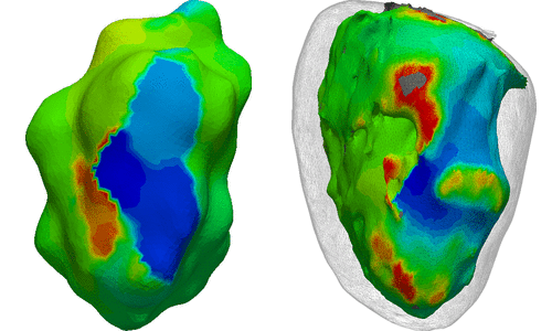
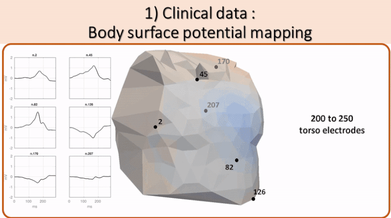 Non invasive cardiac personalisation
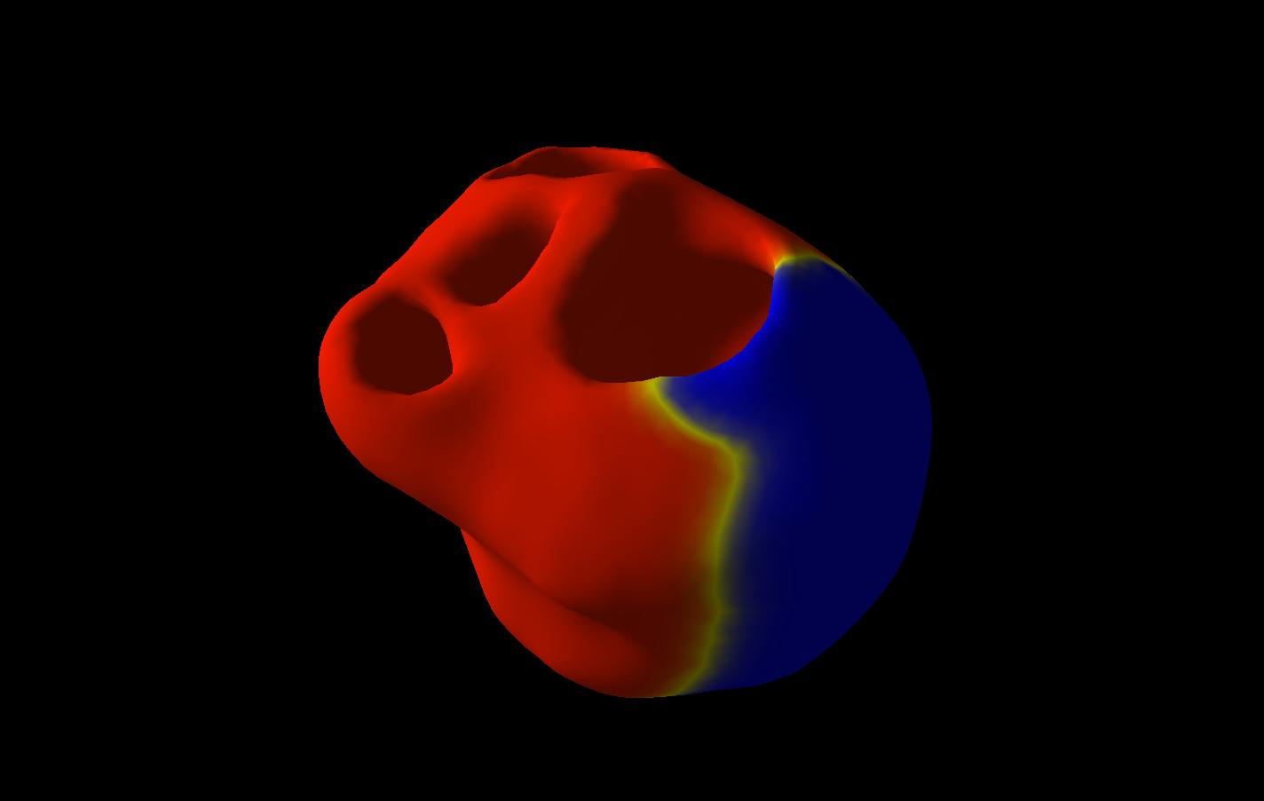
 Simulation of ventricular tachycardia re-entry circuit
 heartMeshFine
 Electrophysiology
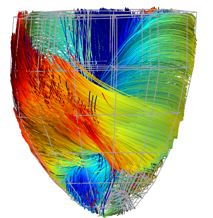 Cardiac Fibres from in vivo Diffusion Tensor Imaging